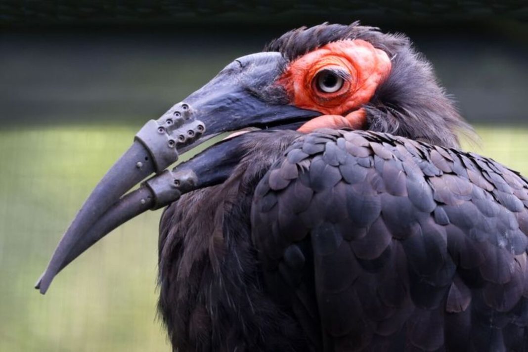

A southern ground hornbill in the Netherlands is making headlines after receiving two custom-made, 3D-printed beak prostheses in a remarkable veterinary breakthrough. The bird at ARTIS Zoo in Amsterdam had damaged both its upper and lower beaks, making everyday activities such as eating and grooming more difficult. Veterinarians and technology specialists joined forces to create replacements tailored to its anatomy. The hornbill began using its new beak shortly after surgery, offering hope for similar treatments in injured birds.
A Southern Ground Hornbill Makes History With Two 3D-Printed Beak Prostheses
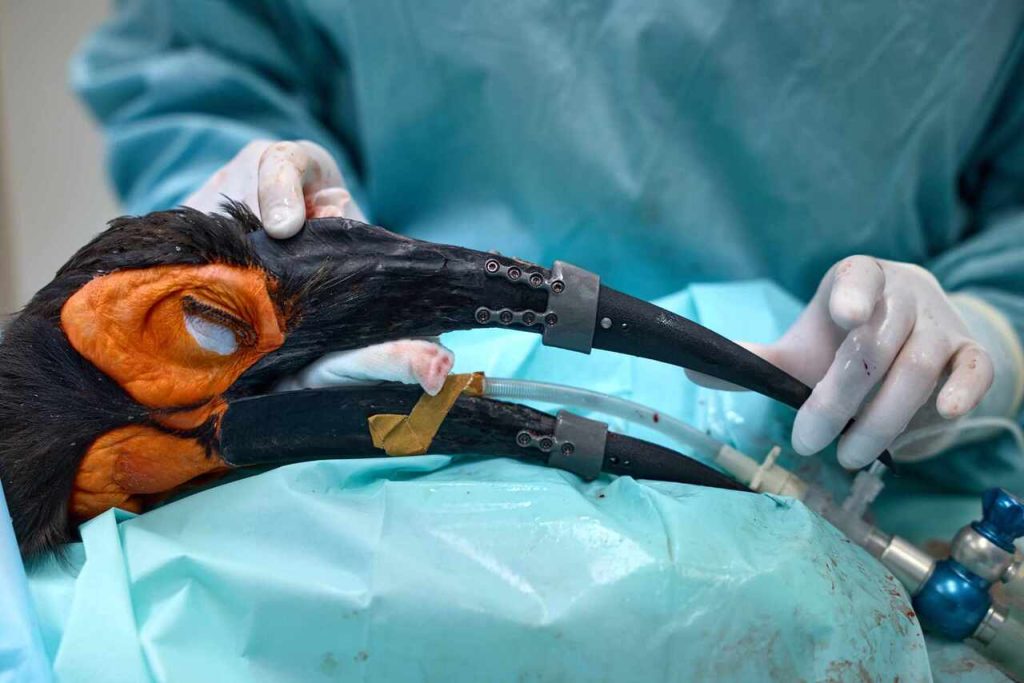
A remarkable veterinary breakthrough in the Netherlands is helping an injured bird regain an essential part of its daily life. A southern ground hornbill at ARTIS Zoo in Amsterdam received two custom-made, 3D-printed prostheses to replace the damaged tips of its upper and lower beaks. According to the zoo, this is the first known case in which both sections of a bird’s beak have been restored using this approach. Although birds have received individual beak prostheses before, replacing both parts presented a more complex challenge because the two sections must fit together and align correctly. The treatment was developed through a collaboration involving ARTIS, Utrecht University’s Faculty of Veterinary Medicine, UMC Utrecht, and medical technology company Amnovis. The team designed the prostheses around the bird’s anatomy and attached them using titanium components and screws. The goal was not simply to restore the hornbill’s appearance, but to improve its ability to perform natural behaviors, including feeding and grooming. The successful early results offer a promising example of how customized medical technology can help veterinarians address injuries that would otherwise limit an animal’s quality of life.
Damage to Both Beak Sections Made Eating and Grooming Difficult
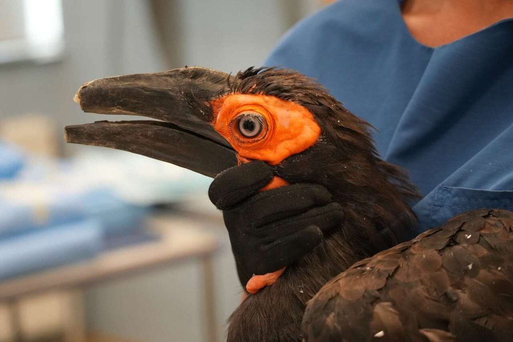
For a bird, a damaged beak can affect far more than the ability to pick up food. The southern ground hornbill at ARTIS Zoo had broken both its upper and lower beaks, leaving two shortened remnants that restricted several everyday activities. Although the bird could manage to some extent, eating became more difficult, particularly when it needed to pick food up from the ground. Grooming was another challenge because birds use their beaks to arrange and clean their feathers, a behavior known as preening. Southern ground hornbills also rely heavily on their beaks while searching for food, including probing and striking the ground. The damage therefore interfered with activities that are central to their normal behavior. Zoo veterinarians wanted to find a solution because the bird was still young and could potentially live for many years. Leaving the injury untreated was one option, but the team believed a carefully designed prosthesis might offer a better long-term outcome. Their decision focused on improving the bird’s welfare by restoring useful function, rather than simply correcting the visible damage.
CT Scans and Original Beak Fragments Helped Create a Custom Fit
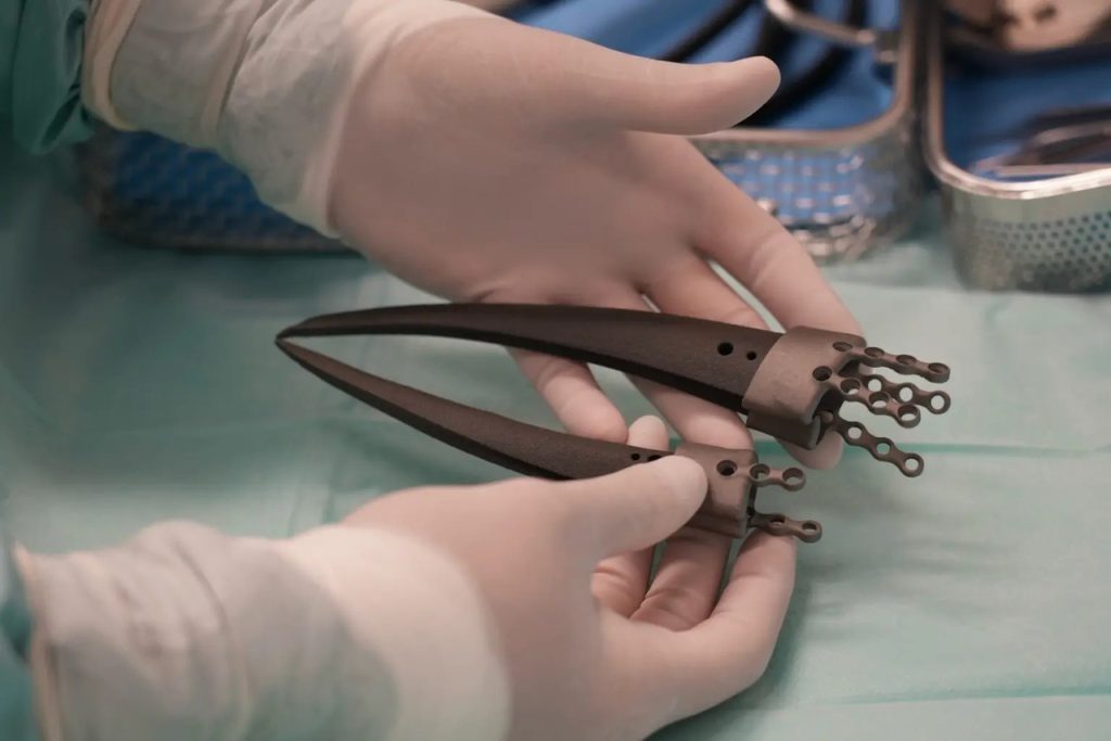
Creating a replacement beak required specialists to reconstruct the bird’s original anatomy with considerable precision. They used a CT scan of its head alongside preserved fragments of the original beak to determine the shape and dimensions needed for the prostheses. This information was important because a bird’s beak must align properly with its skull and with the opposing beak section. Even a small mismatch could make gripping food or performing other movements difficult. Using the scan and original fragments, the team developed a design tailored to the individual hornbill rather than relying on a standard replacement. The prostheses were 3D-printed from plastic material, while titanium components and screws provided the means to attach them securely. The work involved veterinary specialists and technical experts who combined knowledge of bird anatomy with digital modeling and manufacturing. This kind of customization is one of the major advantages of 3D printing in veterinary medicine: it allows replacement parts to be shaped around an animal’s particular needs. The hornbill’s recovery will help the team assess how well the design performs over time.
ARTIS Zoo and Utrecht University Joined Forces for the Procedure
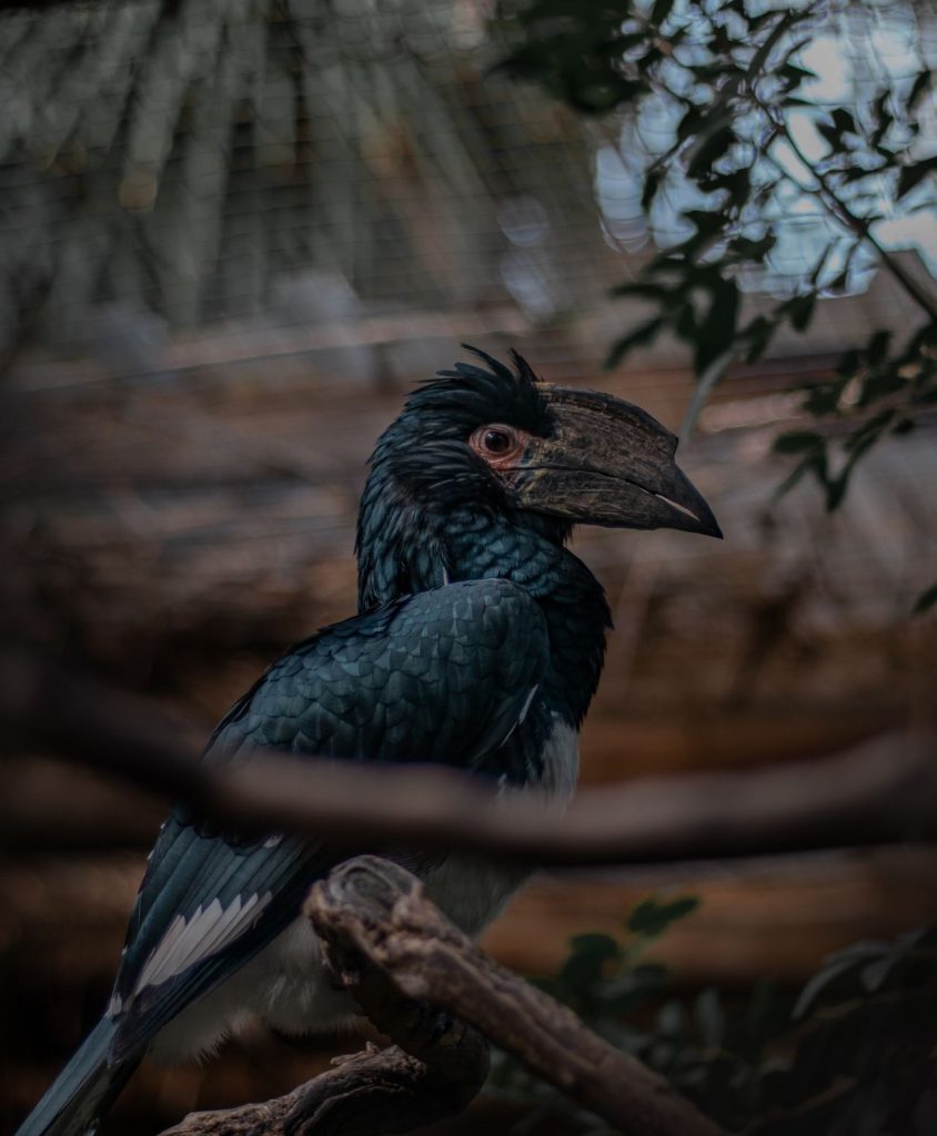
The hornbill’s treatment depended on a team that brought together animal care, veterinary surgery, medical research, and specialist manufacturing. ARTIS Zoo worked with Utrecht University’s Faculty of Veterinary Medicine, UMC Utrecht, and Amnovis to develop and deliver the two prostheses. Veterinarians and bird specialists at ARTIS helped identify the problem and develop a treatment plan focused on the animal’s welfare. Veterinary surgeons from Utrecht University’s Faculty of Veterinary Medicine performed the operation, while UMC Utrecht contributed expertise in designing the replacement beak sections. Amnovis manufactured the titanium components used to secure the prostheses. Each partner had a distinct role, from understanding how the bird used its beak to designing parts that could be attached and function as intended. This collaboration shows why complex veterinary procedures can require expertise beyond a traditional animal hospital. A successful implant must be anatomically suitable, mechanically secure, and practical for the animal to use. The team also needs to monitor the bird after surgery to determine whether the design remains effective. If the prostheses continue to perform well, the experience could provide useful knowledge for veterinarians treating similar injuries in other birds.
The Hornbill Started Using Its New Beak on the Day of Surgery
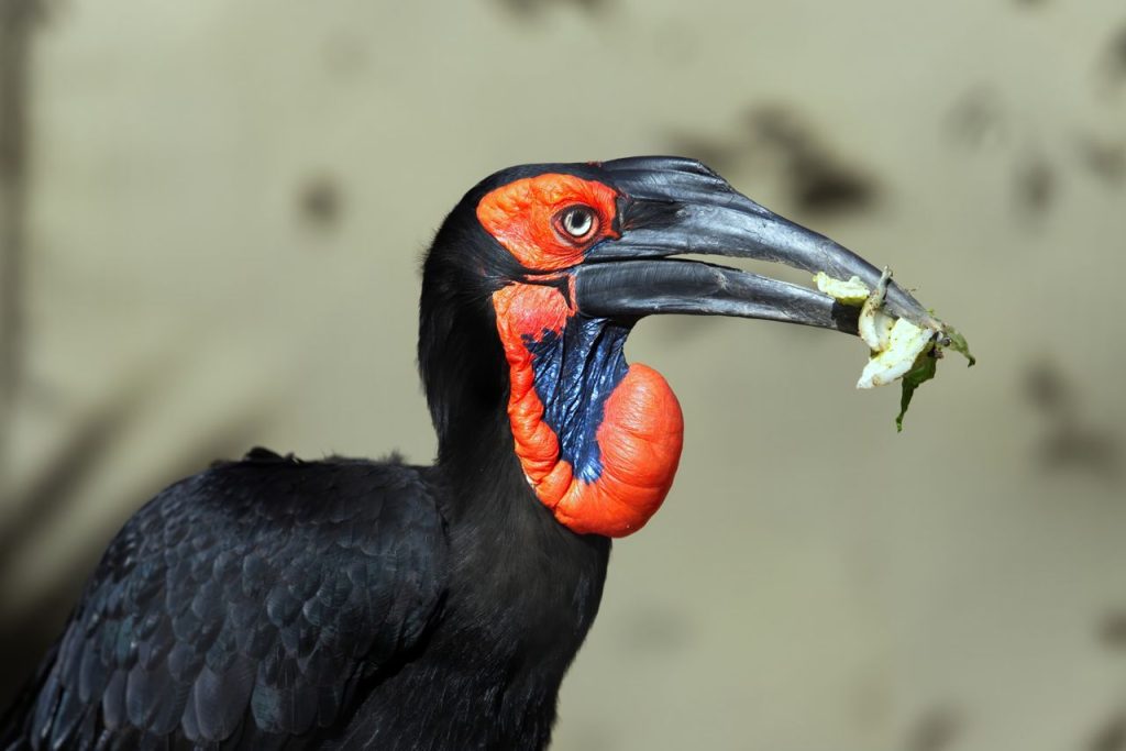
The most encouraging sign came sooner than the veterinary team might have expected. According to ARTIS Zoo, the hornbill began using its new beak on the afternoon of the operation, suggesting that it was adapting to the replacement parts quickly. The next morning, it was able to pick up a small mouse from the ground, and it ate independently later that day. These were important early milestones because the bird’s original injury had made collecting and handling food more difficult. The team had been concerned that the hornbill might reject the prostheses because they feel different from natural beak tissue and do not provide the same sensation. The new parts also added weight and changed the shape of the bird’s beak, meaning it would need time to learn how to use them comfortably. Its early willingness to feed was a positive sign, although it does not guarantee that the prostheses will work perfectly over the long term. Zoo specialists are continuing to monitor the bird’s recovery and behavior. In the days following surgery, it also began preening its feathers, another encouraging indication that the replacement beaks were helping restore natural activities.
Removable Prostheses and Spare Parts Help Prepare for Possible Problems
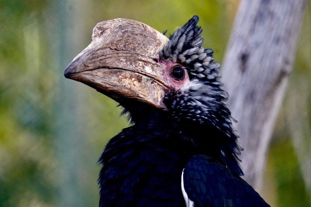
Even a promising medical innovation needs a backup plan, particularly when an animal must live with an artificial replacement. The hornbill’s prostheses were designed with screws so that specialists could remove them if necessary. This precaution mattered because the team could not be certain in advance that the bird would accept the new beak or continue eating normally with it. If the prostheses caused problems, veterinarians needed a way to take them off rather than leave the bird struggling with an unsuitable device. The design also had to account for the fact that the artificial material would feel and weigh differently from the original beak. Although the hornbill began using the replacements quickly, its long-term recovery remains an important part of the project. Specialists will need to observe how the parts hold up during feeding, grooming, and other natural behaviors. The treatment could eventually help other injured birds, but it will not automatically suit every species. Beak shapes, attachment points, and everyday functions differ widely among birds, so each case would require careful assessment. If the prostheses prove durable and effective, the team hopes its experience can contribute to future veterinary research and treatment options.